COMAT 2026
Jun 15, 2026
The 9th annual COMAT 2026 international conference—Modern Trends in Structural Materials—will take place September 8–10, 2026, in Pilsen. ... more
The metallographic laboratory is testing laboratory no. 1476 accredited by the Czech Accreditation Institute:
 |
 |
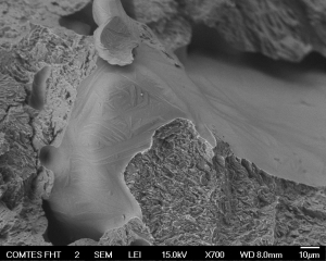
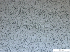
| Microstructure of quenched and tempered steel etched to reveal prior austenite grain boundaries |
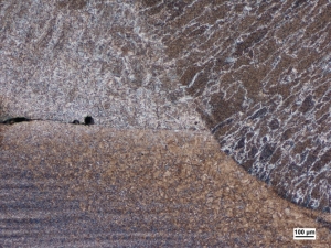
Close-up view of weld |
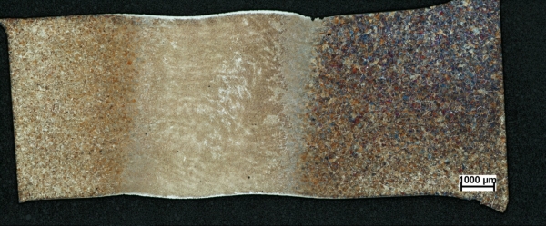 |
| Longitudinal section through a specimen after physical simulation of thermomechanical treatment. Specimen length: approx. 15 mm; specimen width: 6 mm. This micrograph was taken using an optical microscope with an automatic stage and pasted together from 28 fields of view. |
The 9th annual COMAT 2026 international conference—Modern Trends in Structural Materials—will take place September 8–10, 2026, in Pilsen. ... more
From June 1 to 3, 2026, scientists, engineers, and industry representatives from around the world gathered in Pilsen for the 10th annual CHS² - International Conference on Hot Sheet Metal Forming of High-Performance Steel. ... more
Copyright © 2026 COMTES FHT a.s. All rights reserved.
Websites from
![]()